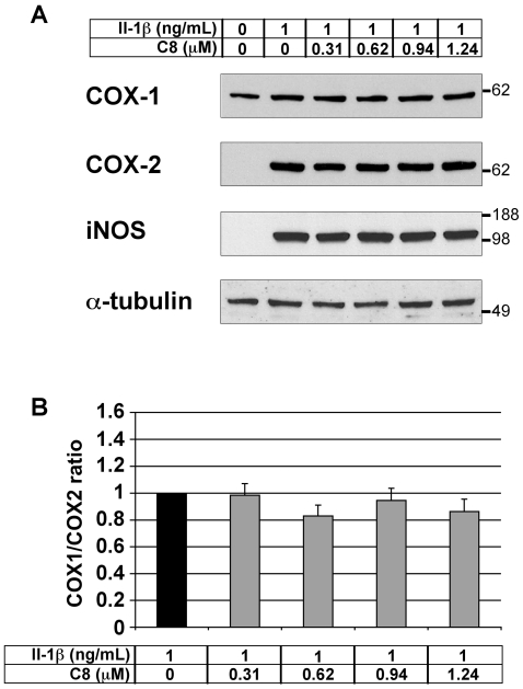
Figure 6. Effect of IL-1β and C8 on the COX-1, COX-2, and iNOS protein levels in articular chondrocytes.
(A) Chondrocytes were untreated or treated for 20 h with IL-1β alone or 1 h after the addition of C8 in DMEM. 20 µg aliquots of whole-cell protein extracts were examined by western blot analysis with antibodies against COX-1, COX-2, and iNOS. α-tubulin immunodetection is shown as a control for protein loading and transfer. Results from one representative experiment in five are shown. (B) Intensities of the COX-1 and COX-2 immunoreactive bands evaluated by semi-quantitative scanning densitometry. Data represent the COX-1/COX-2 protein ratio and are expressed as relative arbitrary units, where the IL-1β-treated group represents 1. Values are means ± SEM (n = 5 independent determinations). No significant differences were found between the groups.

