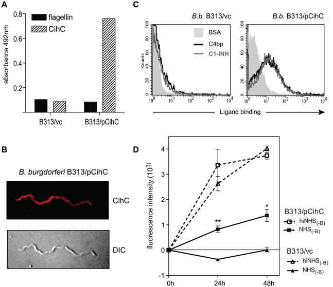
Figure 8. Ectopic expression of CihC in serum-sensitive B. burgdorferi B313.
A) Expression and surface localization of CihC by transformed B. burgdorferi B313 was analyzed by whole cell ELISA using CihC-specific mAb BR2 and as control, the flagellin-specific mAb LA21. B) Binding of C4bp and C1-Inh to B313/vc and transformed B313/CihC cells was analyzed by FACS analysis. C) Immunfluorescence analysis using CihC-specific mAb BR2 followed by rabbit anti-mouse Cy3-conjugated IgG. The corresponding differential interference contrast image is shown in the lower panel. D) For human serum susceptibility assay, mock-transformed B. burgdorferi B313 (B313/vc) and B. burgdorferi B313 transformed with the cihC gene (B313/pCihC) were incubated in the presence of 50% factor-B depleted human serum (NHS-B) or heat-inactivated factor B-depleted human (hiNHS-B) serum at 30°C for 48 h. Cells were stained with a nucleic acid dye and the growth as compared to day 0 was determined by measuring of the fluorescence intensity at 530 nm. Values represent the mean ± SEM of a single experiment performed in triplicate that is representative of three independent experiments. **, P = 0.001; *, P<0.01 for B313/pCihC NHS(-B) at 24h and 48h, respectively, compared to B313/vc NHS(-B).

