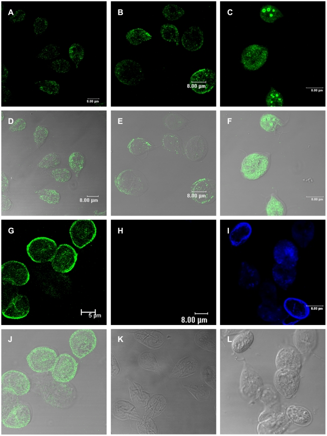
Figure 3. Immunolocalization of Rab11 and CWP1 during encystation of Giardia lamblia.
Samples of trophozoites (A) and parasites of the different cell populations induced to undergo encystation (B, C and G) were processed for confocal microscopy following the protocol described in the Methods section. Images in A–C and G show cells stained with the anti-Rab11 antibody and a FITC-conjugated goat-anti rat secondary antibody. Images in H and I show trophozoites and cells induced to undergo encystation stained with the anti-CWP1 antibody and a Cy5-conjugated goat-anti mouse secondary antibody. D–F and J show superimposed images of cells stained with anti-Rab11 and corresponding DIC images. DIC images in K and L show trophozoites and cysts.

