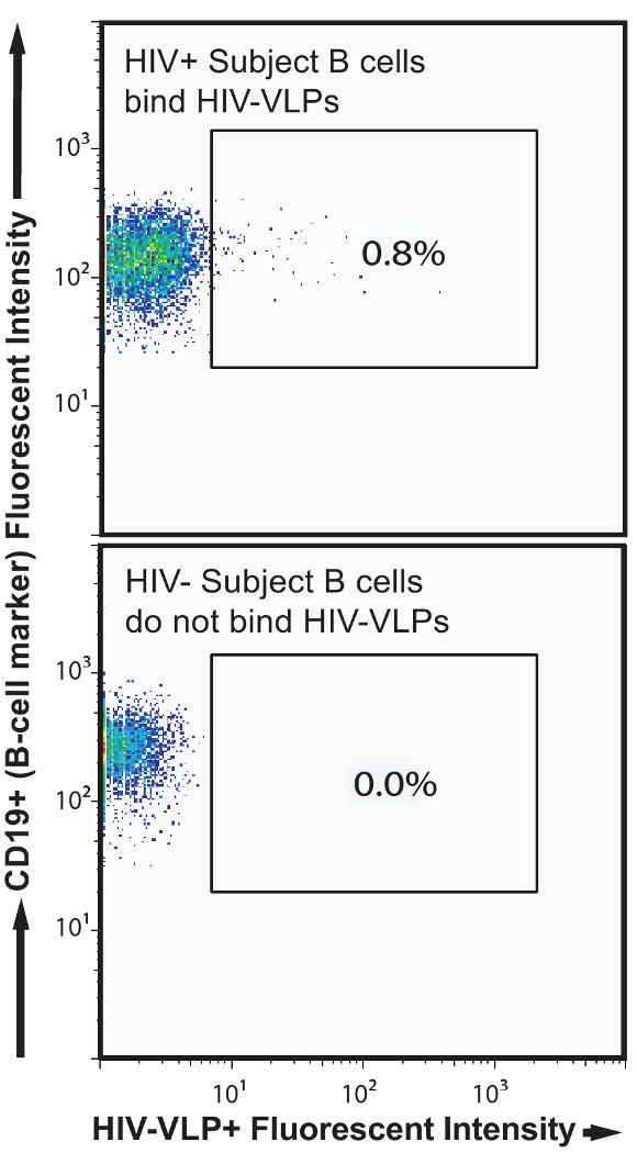
Figure 2. Flow cytometric identification of HIV-specific B cells.

After paramagnetic bead enrichment of B cells from PBMCs, cells from an HIV+ subject (top flow panel) or an HIV- subject (bottom flow panel) were labeled separately with HIV-VLPs and appropriate cell markers. Detection of the anti-CD19 and HIV-VLP reagents are shown after prior gating (not shown) to exclude dead cells, CD3+ T cells, and CD14+ monocytes.
