
Figure 3. Complex binding patterns of antibodies to HIV antigens.
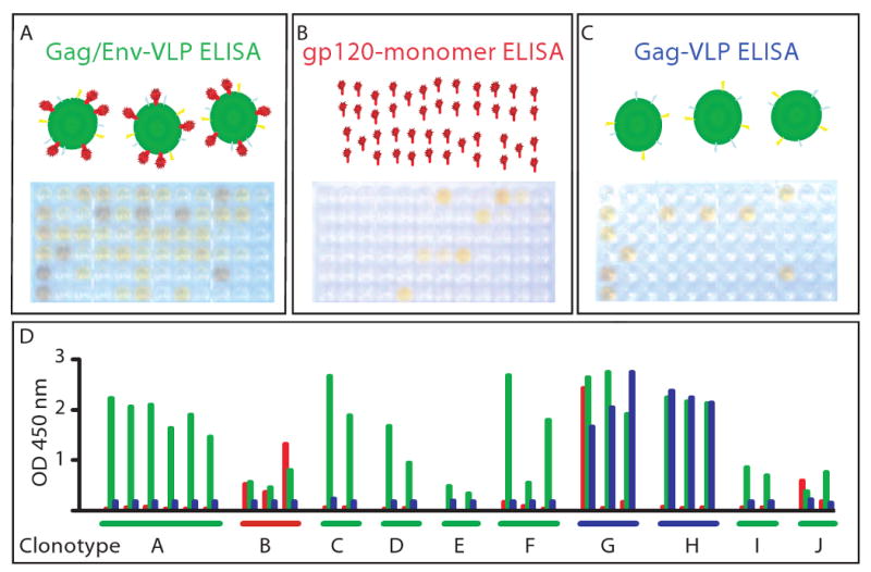
Binding of HIV-specific B cell culture supernates in enzyme linked immunosorbent assay to (A) HIV-VLPs containing Env protein, (B) monomeric gp120, or (C) VLPs containing Gag but not Env protein. Panels A through C show photographs of the colorimetric readout of the reactivity in plates tested with the same supernates arrayed in the same pattern. In (D) is shown the optical density (OD at 450 nm) of the same samples color-coded for reactivity as in the title of the ELISAs above (A=green, B=red, C=blue). In D, the wells are grouped by genetic clonotypes, determined by subsequent sequence analysis of the antibody genes of B cells from corresponding wells. The data in D show that cells within a clonotype (i.e., B cells sharing antibody genes) exhibit similar binding specificity for monomeric (red) or oligomeric (green) forms of Env. Interestingly, two clonotypes (designated G and H) exhibit binding to VLPs that do not contain Env.