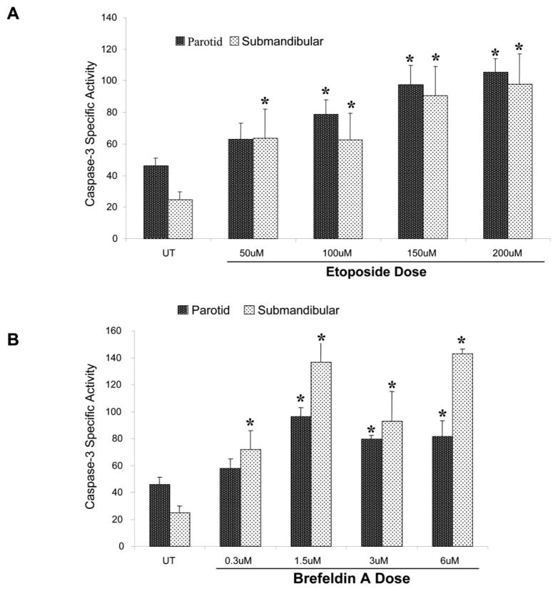
Fig. 1.
Induction of apoptosis in primary cultures of rat parotid and submandibular acinar cells by etoposide and brefeldin A (BFA). Subconfluent primary cultures of parotid and submandibular acinar cells were stimulated with increasing concentrations of etoposide (A) or BFA (B) for 24 h. The concentrations of etoposide and BFA are indicated under each column. All adherent and nonadherent cells were collected and lysed in caspase lysis buffer (BioMol QuantiZyme Colormetric Assay kit). Fifteen micrograms of cell lysate was used to analyze the level of enzyme-specific activity (pmol/min/μg protein) for each sample, and samples were measured in quadruplicate as described in the Materials and Methods. The results obtained with the primary parotid cells are indicated in black columns, whereas the results obtained with the primary submandibular cells are indicated in the stippled columns. Error bars represent standard error of the mean from four independent experiments. Student’s t-test P values were calculated using Microsoft Excel, and asterisks designate statistical significance (P ≤ 0.1) from the respective untreated control.

