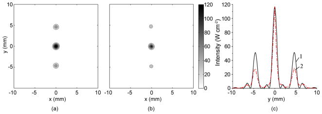
Figure 13.
Intensity distributions in the focal plane (z=130 mm) for the partially activated array for a single focus located at (0, 0, 130 mm) without a rib phantom placed in the beam path. (a) predicted intensity distribution; (b) IR measured intensity, (c) 1D distribution of the corresponding quantities over the vertical coordinate y perpendicular to the direction of ribs for theory (curve 1) and experiment (curve 2). The contours in (a,b) are given with increments of 20 W cm−2. Measurements and simulations were carried out for the acoustic power of 11 W and time of heating of 0.2 s.

