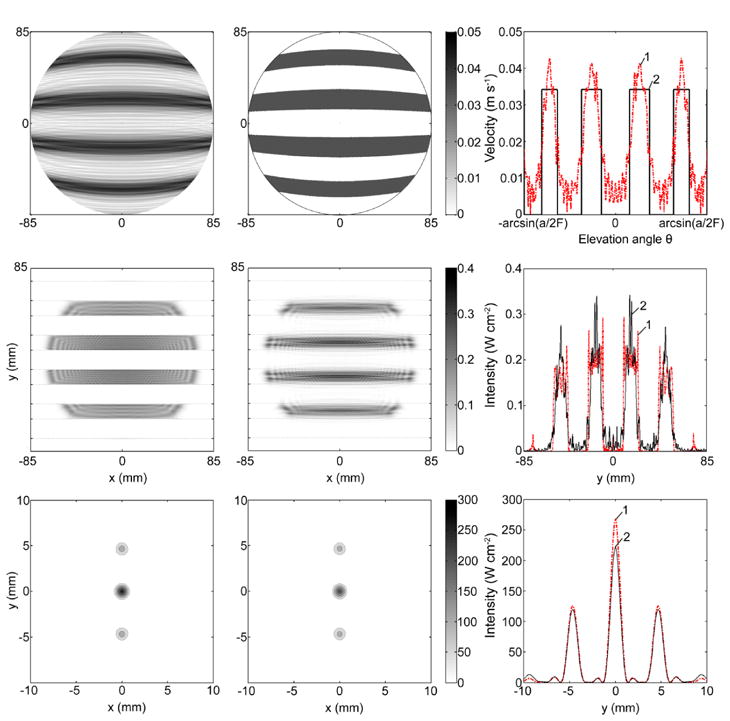
Figure 6.

Modeling results for an idealized radiator and infinitely thin fully absorbing ribs. The velocity distribution at the radiator surface (upper row), the intensity distribution in the plane of ribs (central row) and in the focal plane (lower row) obtained using the diffraction (left column) and geometric approaches (central column). The right hand column shows 1D distributions of the corresponding values over the vertical coordinate y perpendicular to the direction of ribs for diffraction (dashed curve 1) and geometric (solid curve 2) approaches.
