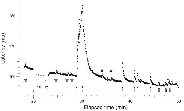
Figure 5.
Record from the superficial peroneal nerve of a patient with painful neuropathy (#16, Table 1). This fiber with a conduction velocity of 0.59 m.s−1, had a latency increase of 0.5% after the 1/30 Hz pause, typical of a CMH. It displayed abnormal discharges (gray arrows) detected as activity-dependent conduction slowing equivalent to one or two action potential spikes. This was determined according to a calibration after the 2-Hz tetanus when 3 extra electrical stimuli were given during the ongoing 0.25 Hz stimulation to demonstrate the extent of activity-dependent slowing (asterisks). This unit was abnormally sensitive to mechanical stimulation (threshold 7.8 mN) (solid arrows).

