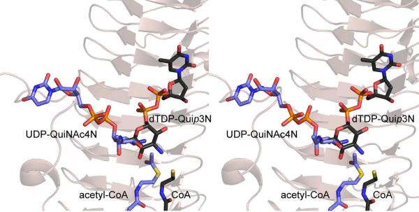
Figure 1.
Differences in the orientations of the nucleotide-linked sugar substrates when bound to PglD or QdtC. The ribbon representation in the background corresponds to that for QdtC. The substrates for PglD and QdtC are highlighted in blue and black bonds, respectively. All figures were prepared with the software package PyMOL (31)

