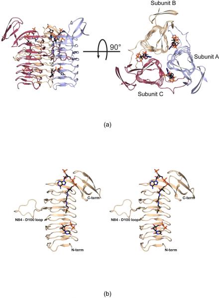
Figure 2.
The structure of WlbB complexed with acetyl-CoA and UDP. A ribbon representation of the WlbB trimer is shown in (a) with the three subunits displayed in red, wheat, and light blue and the bound ligands shown as sticks. The overall three-dimensional fold of one subunit is presented in stereo in (b). The loop defined by Asn 84 to Asp 100 interrupts the regularity of the LβH motif.

