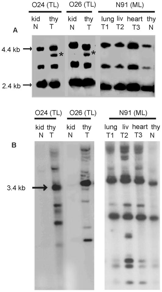
Figure 1.
Southern blot analysis of genomic DNA from tumor (T) or paired normal (N) tissue from FeLV-infected cats O24, O26 and N91 bearing thymic lymphoma (TL) or multicentric lymphoma (ML) (Chandhasin et al., 2005). A. DNA samples (8 μg) were digested with KpnI and hybridized to probe B/S, a Sau3A fragment from the env gene of FeLV-B/Gardner-Arnstein specific for the major classes of endogenous FeLV that serve as substrates for recombination. The distinctive hybridizing fragment of ~3.6-kb (*) indicates recombinant FeLV-B provirus in genomic DNA (Chandhasin et al., 2005; Tsatsanis et al., 1994). B. KpnI-digested DNA samples were also hybridized to a probe representing the U3 region of the LTR of exogenous FeLV. By this analysis, clonally integrated proviruses are visualized as host-virus junction fragments in tumor DNA (Levy, Gardner, and Casey, 1984).

