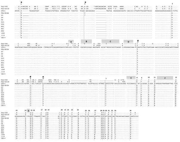
Figure 3.
Nucleotide sequence of the PCR amplification products from tumor DNA of the indicated animals aligned with the sequence of endogenous FeLV-related provirus CFE-6. The previously reported sequences of FeLV-945, FeLV-A/61E and FeLV-B/Gardner-Arnstein are aligned for comparison. Nucleotide sequence differences as compared to CFE-6 are indicated; (…) indicates sequence identity with CFE-6; (____) indicates deletion relative to CFE-6; (//) indicates a break in the sequence shown. Indicated above the sequence are the positions of 34 nucleotide sequence differences in the amplification products as compared to CFE-6. Those indicated with an asterisk (*) result in amino acid change relative to CFE-6. Also indicated about the sequence are recombination sites A – G, previously reported to be preferred sites for crossover in the generation of FeLV-B (Sheets et al., 1992).

