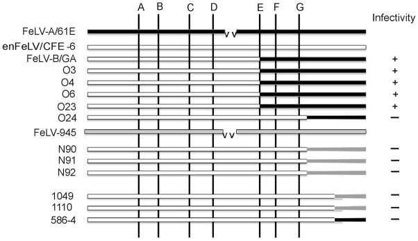
Figure 4.
Schematic representation of the recombination junctions identified in the SU genes of FeLV-B viruses amplified from tumor DNA of the indicated animals. Depicted for comparison are the SU genes of exogenous viruses FeLV-A/61E (solid bar) and FeLV-945 (gray bar) and endogenous FeLV-related provirus CFE-6 (open bar). A deletion within FeLV-A/61E relative to CFE-6 is indicated (vv). Shown diagrammatically are the positions of previously identified preferred recombination sites A – G (Sheets et al., 1992). Also indicated is the infectivity of genomic DNA for canine-D17 cells, presumably representing polytropic FeLV-B replication.

