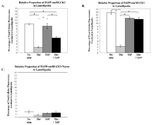
Figure 2. Relative proportion of nmMLCK isoforms in human lung endothelial lamellipodia.
Human lung EC were treated as described in Figure 1, and the percentage of total spatially–specific cellular distribution of nmMLCK isoforms determined (see Methods) after vehicle, thrombin for 10 min, S1P for 30 min, or thrombin for 10 min followed by S1P for 30 min. Panel A: The percentage of total cellular EGFP-nmMLCK1 localized to lamellipodia is indicated for each condition. * p<0.05; N = 4–18 per condition. Panel B: The percentage of total cellular EGFP-nmMLCK2 localized to lamellipodia is indicated for each condition. * p<0.05; N = 7–17 per condition. Panel C: The percentage of total cellular EGFP-nmMLCK2Nterm localized to lamellipodia is indicated for each condition. Less than 2% is localized to lamellipodia in each condition. N = 10–14 per condition.

