Figure 8.
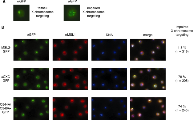
In vivo localization of different MSL2 derivatives. Stable Drosophila SL2 cell lines, which express different MSL2 versions fused to GFP, were analyzed by immunofluorescence staining. Localization of MSL2-GFP derivatives were visualized using anti-GFP antibodies (α-GFP). Endogenous MSL1 was detected using anti-MSL1 antibodies (α-MSL1). DNA was counterstained with Hoechst 33258 (DNA). (A) Examples showing single nuclei with proper targeting of MSL2 to the X chromosomal territory (left) or dispersed, non-physiological distribution of the GFP fusion proteins. (B) Representative fields of cells. Cells that express the MSL2-GFP transgene were analyzed for proper X territory staining. The percentage of cells, which show mislocalized, dispersed GFP signals from the total number of GFP positive cells counted are displayed to the right.

