Fig. 4.
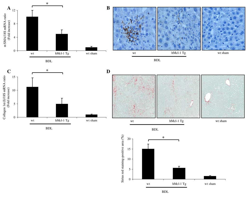
Hepatic fibrosis is reduced in hMcl-1 Tg animals 14 days post-BDL. a α-SMA and collagen 1α (I) mRNA expression, markers for stellate cell activation and hepatic fibrogenesis were quantified by real time-PCR. Data were obtained from ten independent animals and expressed as mean ± standard error (*P < 0.05 by ANOVA). b Photomicrographs after immunohistochemistry for α-SMA following BDL of 14 days are depicted. c Expression of collagen 1α (I) mRNA was quantified by real time-PCR 14 days after BDL (*P < 0.05 by ANOVA, n = 10 for each group). d Sirius red staining, a chemical stain of collagen deposition in the liver, was performed 14 days after BDL. Collagen fibers stained with Sirius red were quantitated using digital image analysis. Representative photomicrographs of liver sections from each mouse strain are depicted (magnification by light microscopy 40×; *P < 0.05 by ANOVA, n = 10 for each group)

