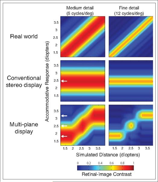
Fig. 4.
Retinal-image contrasts for different display techniques. In each panel, simulated distance (in diopters) is plotted as a function of the distance to which the eye accommodates (in diopters). Retinal-image contrast for an object of contrast 1 is indicated by the colors. The white arrows in the middle and bottom rows represent the distances to the image planes.

