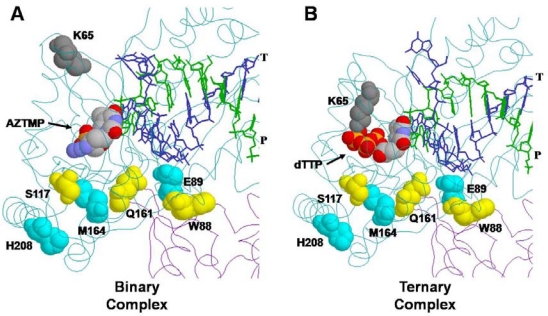
Figure 2.
Locations of residues altered by PFA-resistance mutations in the structures of binary and ternary complexes of HIV-1 RT. (A) Binary complex of HIV-1 RT with AZTMP-terminated primer-template occupying the dNTP-binding site (N site) [PDB structure 1N6Q, Ref. 45]. (B) Ternary complex of HIV-1 RT with ddAMP-terminated primer-template and dTTP occupying the N site [PDB structure 1RTD, Ref. 85]. The template (T) is shown in blue, and the primer strand (P) in green. The structure occupying the N site in each complex is shown as a space-filling model (atoms indicated by CPK color scheme). Residues that are substituted in the indicated PFA-resistant mutants are also shown as space-filling models. The fingers-domain residue K65 is shown in gray. Palm-domain residues are shown in yellow or cyan using contrasting colors to distinguish overlapping structures [37].

