
Figure 1. Structure of the sIL-10R2 chain.
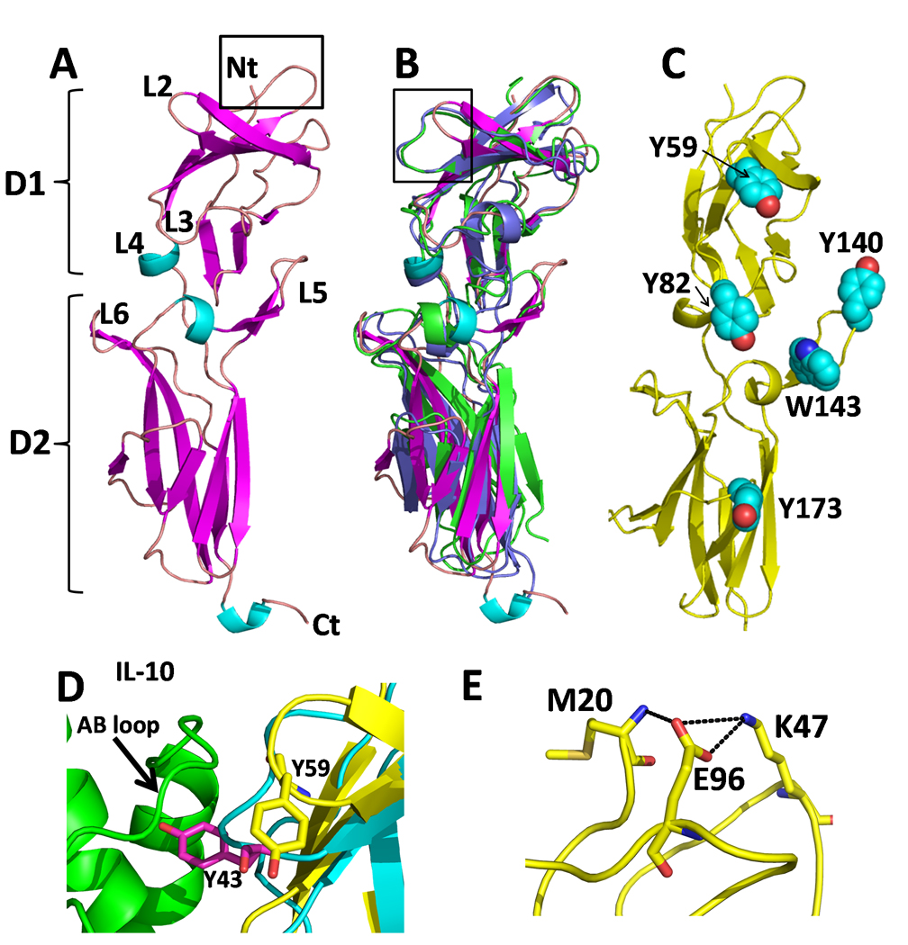
(A) Ribbon diagram of the sIL-10R2 chain colored by secondary structure with binding loops labeled. Box shows the location of Figure F. (B) Superposition of sIL-10R2 (colored as in A) with sIL-10R1 (green) and sIL-22R1 (purple). Box shows the location of Figure D. (C) Location of aromatic residues on sIL-10R2 on sIL-10R2. (D) Comparison of the high affinity site 1 interaction between sIL-10R1 Y43sIL-10R1 and the AB loop of IL-10 (green) with the sIL-10R2 L2 loop and Y59sIL-10R2 (yellow). (E) Interaction network for K47sIL-10R2. Replacement of K47sIL-10R2 with a glutamic acid is associated with persistent HBV infection (Frodsham et al., 2006).