
Figure 2. Unique clefts identified in sIL-10R2.
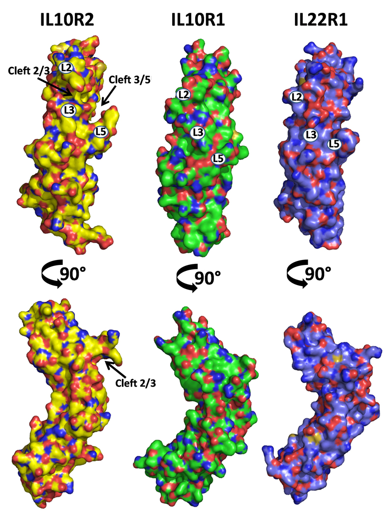
The structures of sIL-10R2, sIL-10R1, and sIL-22R1 are shown as molecular surfaces. Images in the top row are orientated as found in Figure 1A. The surfaces are colored by atom type with oxygens red, nitrogens blue, and sulfur orange. Carbons are colored yellow, green, and purple in sIL-10R2, sIL-10R1, and sIL-22R1, respectively. Numbers on the surfaces correspond to loop positions.