Figure 1.
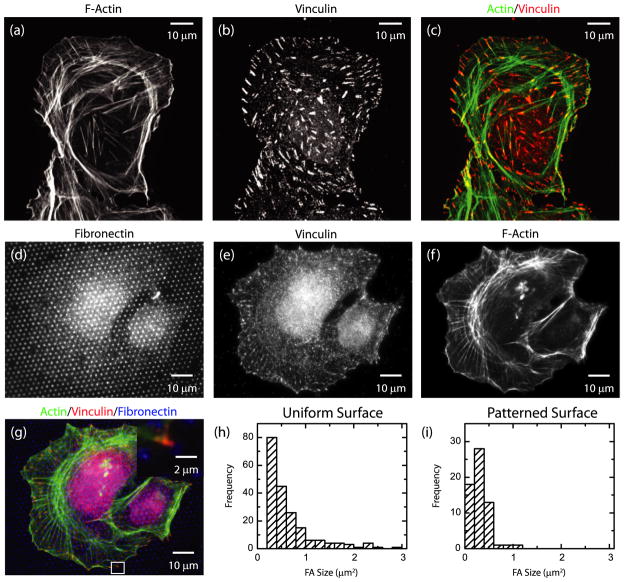
(a)–(c) Immunofluorescence image of a U2OS cell plated on a substrate uniformly coated with fibronectin. (a) F-actin; (b) vinculin; (c) color combine with F-actin in green and vinculin in red. (d)–(g) Immunofluorescence image of a U2OS cell plated on a substrate micro-patterned with fibronectin. (d) Fibronectin; (e) vinculin; (f) F-actin; (g) color combine with actin in green, vinculin in red, fibronectin in blue. Inset: magnified image of region indicated by white box. (h) Histogram of focal adhesion areas on a uniform substrate. (i) Histogram of focal adhesion areas on a patterned substrate as in (d).

