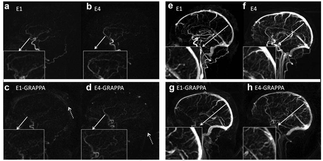
FIG. 7.
Arterial (left) and venous (right) phases of four-dimensional MRA using CAMERA of four volunteers. E4 shows superior image quality during the arterial phase, with better delineation of the ACA and E1, for both GRAPPA and non-GRAPPA versions. More smaller vessels are identified near the sagittal sinus during the venous phase with E4 than E1 acquisitions, both with and without GRAPPA.

