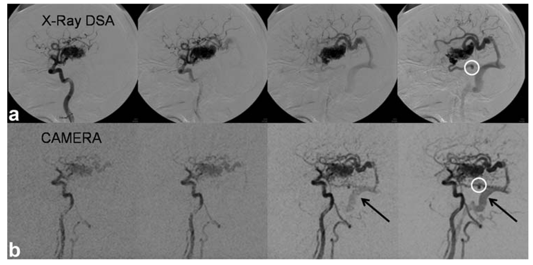
FIG. 8.
AVM patient image acquired with E4 (inverted). Comparison with X-ray digital subtraction angiography images show close correlation. Early drainage from the AVM to the sigmoid sinus is clearly depicted (black arrows). A small aneurysm (white circle) is also visible with both X-ray and MRA. Temporal footprint = 8 sec.

