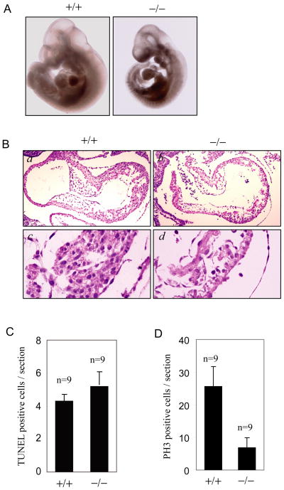
Figure 1. SENP2−/− embryos have a defect in myocardial development.
(A) Appearance of SENP2+/+ and SENP2−/− embryos at E10.5. The SENP2−/− embryo has a smaller heart with pericardial effusion.
(B) H& E-stained sections of heart from E10.5 wildtype (a, c) and SENP2−/− (b, d) embryos. The SENP2−/− embryos showed hypocellular endocardial cushions and myocardial hypoplasia with thinner myocardium.
(C) TUNEL staining of sections of hearts from SENP2 +/+ or −/− embryos at E10.5 showed no significantly increased in apoptosis in the mutant embryos (P>0.01). “n” indicates the number of sections examined.
(D) Phosphohistone H3 (PH3) staining of sections of hearts from SENP2 +/+ or −/− embryos at E10.5 revealed reduced proliferation in the mutant embryos (P<0.01). “n” indicates the number of sections examined.

