Figure 7.
Effects of G2/M Delay on dCel1p Level.
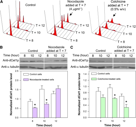
(A) Flow cytograms of PI-stained synchronous C. cohnii cells harvested at different time points after cell cycle synchronization. Microtubule inhibitors, nocodazole and colchicine, delayed the cell cycle at the G2/M phase. The arrows indicate the G2/M delay (the second peak was higher than the first peak at T = 12 when compared with the control) upon nocodazole and colchicine treatments.
(B) and (C) Immunodetection of dCel1p in cells treated with nocodazole and colchicine, respectively. Anti-α-tubulin was used as a loading control. Immunodetectable signals for dCel1p were determined using ImageJ and normalized to the corresponding α-tubulin signals. Blots are representative of three independent experiments. Both nocodazole and colchicines led to a significant decrease (P < 0.05) in the abundance of immunodetectable dCel1p at T = 10 when compared with the control. Data represent means ± se of three replicate experiments. Asterisk indicates significant (P < 0.05) difference compared with the corresponding control.
[See online article for color version of this figure.]

