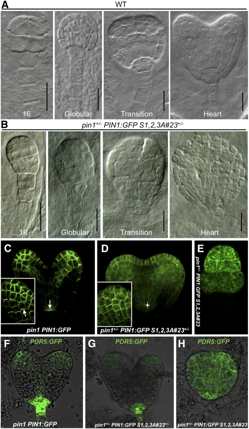
Figure 6.
Embryo Defects Induced by PIN1:GFP S1,2,3A Mislocalization Are Due to Disturbed Auxin Distribution.
(A) and (B) Differential interference contrast microscopy images of embryos from wild-type (A) and pin1+/− PIN1:GFP S1,2,3A#23+/− (B) plants. Text at the bottom of each image indicates the developmental stage of the embryo. For the defective embryos in (B), the developmental stage was based on a rough estimate of the cell number. Bars = 10 μm.
(C) to (E) Confocal laser scanning microscopy images of pin1 PIN1:GFP heart-stage embryos (C) and wild-type looking (D) and defective looking (E) embryos from pin1+/− PIN1:GFP S1,2,3A#23+/− plants from the same developmental stage. Insets in (C) and (D) represent confocal scans through the epidermal cell layer of cotyledon primordia. White arrows in (C) and (D) indicate the PIN1:GFP polarity, and a star in (D) indicates the absence of basally localized PIN1:GFP S1,2,3A protein.
(F) to (H) Confocal laser scanning microscopy images of PDR5:GFP auxin distribution in embryos from pin1 PIN1:GFP (F) and pin1+/− PIN1:GFP S1,2,3A#23+/− ([G] and [H]) plants, showing a reduced (G) or mislocalized (H) auxin maximum.

