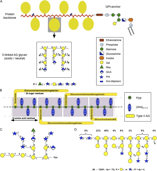
Figure 1.
A, The wattle blossom model of the structure of AGPs with a GPI membrane anchor attached. In this model, there are approximately 25 Hyp residues. Most Hyp residues are noncontiguous and are predicted to bear an AG chain. Each AG chain may contain 15 or more repeats of a β-(1-3)-linked Gal oligosaccharide. There may be a few contiguous Hyp residues bearing short arabino-oligosaccharides. The molecule as a whole is spheroidal. The structure of the GPI anchor shows an ethanolamine-phosphate (P) between the anchor and the C terminus of the protein backbone, which is common to all GPI anchors. The core oligosaccharide of the GPI shown is based on PcAGP1 from pear (Pyrus communis; Oxley and Bacic, 1999), which comprises 2- and 6-linked Manp residues, a 4-linked GlcNH2 residue, and a monosubstituted inositol with a partial Galp residue substitution to C(O)4 of the 6-linked Manp residue. The lipid moiety is a ceramide composed primarily of a phytosphingosine base and tetracosanoic acid. This model is modified from Fincher et al. (1983). B, The twisted hairy rope model of the structure of the GAGP. A hypothetical block size of 7 kD contains 10 amino acid residues (1 kD), 30 sugar residues (4.4 kD), and three Hyp-triarabinosides (1.32 kD). The glucuronorhamnoarabinogalactan has a galactan backbone with GlcpA, Rhap, and Araf side chains similar to that shown in the AG schematic in A. This model is from Qi et al. (1991). C, Primary structure of a representative Hyp-AG polysaccharide (AHP-1) released by base hydrolysis from a synthetic AGP (Ala-Hyp)51 from tobacco BY2 cells. This model is modified from Tan et al. (2004). D, Larch AG structure. R′ = dp > 4 is an undefined AG oligosaccharide. This model is modified from Ponder and Richards (1997).

