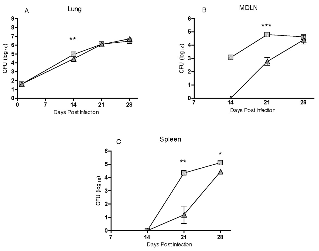
FIGURE 2.
Trafficking of M. tuberculosis from the lungs to the MDLN and spleen is delayed in CCR7−/− mice compared with controls. CCR7+/+ and CCR7−/− mice were sacrificed at the indicated time points after aerosol infection with a low dose of M. tuberculosis and colony-forming units were determined in homogenates of lungs, mediastinal lymph node, and spleen. Data are represented as mean ± SD of four mice per group and per time point. * P< 0.05, ** P<0.005, *** P<0.001 by unpaired Student’s t test.

