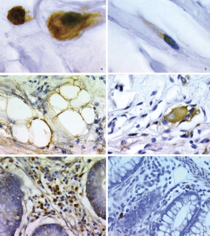
Fig. 11.
Evidence of increased abundance of IGF-IR+ cells in Crohn's disease: a, inflammatory cells; b, fibroblastoid cell; c, adipocytes; d, hypertrophied nerve plexus. Note the relative frequencies of IGF-IR+ cells in the lamina propria in uninvolved (e) and disease-involved (f) areas. [Reproduced from El Yafi F, Winkler R, Delvenne P, Boussif N, Belaiche J, and Louis E (2005) Altered expression of type I insulin-like growth factor receptor in Crohn's disease. Clin Exp Immunol 139:526–533. Copyright © 2005 British Society for Immunology. Used with permission.]

