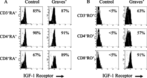
Fig. 7.
Disproportionate IGF-IR+CD45RO+ memory T cells from patients with GD. The fraction of CD3+, CD4+, and CD8+ T lymphocytes expressing IGF-IR was determined using multiparameter flow cytometry by gating on populations of CD3+, CD4+, or CD8+, CD45RA+, or CD45RO+ T cells and is represented as a histogram (filled) compared with isotype controls (open). A, naive CD45RA+ lymphocytes from a patient with GD and a control donor demonstrate a similar, frequent display of IGF-IR. B, the fraction of memory CD45RO+ lymphocytes expressing IGF-IR is dramatically greater in lymphocytes from a patient with GD compared with control. GD CD8+CD45RO+ T lymphocytes uniformly express IGF-IR, compared with infrequent control CD8+CD45RO+ cells. T-cell expression of IGF-IR was representative of our aggregate observations. [Reprinted from Douglas RS, Gianoukakis AG, Kamat S, and Smith TJ (2007) Aberrant expression of the IGF-1 receptor by T cells from patients with Graves' disease may carry functional consequences for disease pathogenesis. J Immunol 178:3281–3287. Copyright © 2007 The American Association of Immunologists, Inc. Used with permission.]

