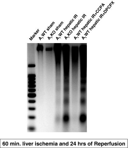
Fig. 7.
Representative gel images (of four experiments) demonstrating DNA laddering as an index of DNA fragmentation in the kidney tissues from sham-operated A1WT (A1WT sham) and A1KO mice (A1KO sham), A1WT (A1WT hepatic IR) or A1KO mice (A1KO hepatic IR) subjected to 60 min of hepatic ischemia and 24 h of reperfusion, and A1WT mice pretreated with 0.4 mg/kg DPCPX (A1WT hepatic IR+DPCPX) or 0.1 mg/kg CCPA (A1WT hepatic IR+CCPA) and subjected to 60 min of hepatic ischemia and 24 h of reperfusion. Apoptotic DNA fragments were extracted according to the methods of Herrmann et al. (1994). This method of DNA extraction selectively isolates apoptotic, fragmented DNA and leaves behind the intact chromatin.

