Fig. 8.
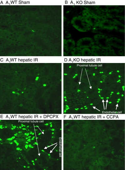
Representative fluorescent photomicrographs (of four experiments) illustrate apoptotic nuclei (TUNEL fluorescent stain; magnification 400×) in kidney sections from sham-operated A1WT mice (A1WT sham; A) and A1KO mice (A1KO sham; B), A1WT mice (A1WT hepatic IR; C) or A1KO mice (A1KO hepatic IR, D) subjected to 60 min of hepatic ischemia and 24 h of reperfusion, and A1WT mice pretreated with 0.4 mg/kg DPCPX (A1WT hepatic IR+DPCPX; E) or 0.1 mg/kg CCPA (A1WT hepatic IR+CCPA; F) and subjected to 60 min of hepatic ischemia and 24 h of reperfusion. In the kidney, endothelial cells predominantly underwent apoptotic death (short, thick arrows) with sparing of renal proximal tubule cells (long, thin arrows) as illustrated in Fig. 6D.

