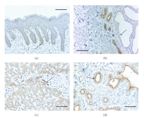
Figure 3.
Immunohistochemical staining of sections of human skin a, and b and liver c, and d with antibodies against PKP 2. (a) The staining of samples of human skin with a monoclonal antibody against PKP 2 (clone PP2-150; Progen, Heidelberg) demonstrates a weak and delicate desmosomal staining as well as cytoplasmic staining in the basal layer of the interfollicular epidermis (arrow). Suprabasal keratinocytes remain unstained. (b) Eccrine sweat glands and ducts show a strong reaction with PKP 2-specific antibodies while apocrine sweat glands exhibit an apical, distinct but weak desmosomal reaction (arrow). (c) Hepatocytes as well as bile ductules are marked at the cell-cell contacts by PKP 2-specific antibodies (arrow). (d) Bile ducts also show a sharp and apical staining of desmosomal structure by the PKP 2-antibodies. The samples shown in (c) and (d) are derived from liver tissue in the vicinity of a metastasis of a gastrointestinal stromal tumor with portal and periportal fibrosis and ductal and ductular proliferation. Scale bars: 100 μm (d), 200 μm (a, b, c).

