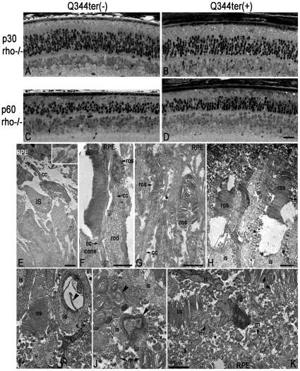
Figure 4. Q344ter transgene does not accelerate retinal degeneration in rho−/− retinas.
As in Figure 2, these images of retinal sections from epoxy-embedded eyecups were taken just above the optic nerve region from Q344terrho−/− (B, D) and their transgene-negative littermate control (A, C) mice at p30 and p60. At both time points, ONL thicknesses appear similar in Q344terrho−/− mice and their littermate controls (compare A to B and C to D). Although progressive ONL thinning was observed in both groups, Q344ter does not appear to accelerate degeneration already occurring in rho−/− mice. Scale bar in D (20 µm) is representative for panels A–D. (E) Rho−/− rod photoreceptors do not elaborate outer segment structures. Instead, membrane tubules are seen (inset). The subretinal space is devoid of vesicular structures. Panels F–H are from p30 Q344terrho−/− retinas, and I–K are from p60 Q344terrho−/− retinas. (F) Short and disorganized rod outer segment can be seen distal to the connecting cilia. A neighboring cone photoreceptor with a much more intact outer segment is shown for comparison. (G) The rod outer segments shown here are thinner than normal (compare with Figure 2D). These structures are surrounded by apical processes from the RPE. (H) The membranous discs within some outer segment structures appear to be unstable. Numerous vesicular structures are present in the extracellular space (asterisks). (I-K) Vesicular structures are present in the interphotoreceptor space of Q344terrho−/− retinas. Outer segments containing discs are evident, but they are significantly compromised both in size and organization. Scale bars for E-K = 1 µm. Panels I-K are taken at the same magnification. os, outer segment; ros, rod outer segment; is, inner segment; cc, connecting cilium; RPE, retinal pigmented epithelium; m, membranous debris.

