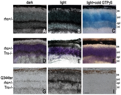
Figure 7. Light-dependent GTPγS loading (20 min exposure) in transgenic Q344ter frozen retinal sections.
[35S]GTPγS binding in situ was performed on unfixed frozen retinal sections from mice with the indicated genetic backgrounds. Basal [35S]GTPγS loading in the dark labels the synaptic layers (A, D, E), while light-exposure lead to additional labeling at the inner and outer segment compartments (B, E, H). Panels C, F, and I show non-specific background labeling. Scale bar = 20 µm. All panels are taken at same magnification.

