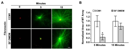
Fig. 1.
Rapid microtubule regrowth in HFFs is inhibited in serum-free DMEM. (A) Representative images of microtubule regrowth in serum-starved HFFs replated onto fibronectin in CCM1 or serum-free (SF) DMEM, where α-tubulin staining is in green and γ-tubulin staining is in red. (B) Quantification of the area of microtubule regrowth. Plotted is the normalized mean area at 5 and 15 minutes post-nocodazole washout ± s.d. calculated from 150 cells per condition in each of eight independent experiments. *Regrowth in SF DMEM is statistically different from regrowth in CCM1 (P<0.05). Scale bar: 2 μm.

