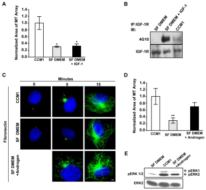
Fig. 2.
Androgen promotes microtubule regrowth. (A,B) IGF1 is not sufficient to promote rapid microtubule regrowth. (A) Microtubule regrowth was compared in serum-starved HFFs replated onto fibronectin in CCM1, SF DMEM and in SF DMEM plus IGF1, with IGF1 only added for the regrowth period. Values are the normalized mean area of microtubule arrays at 5 minutes post-nocodazole washout ± s.d. from 150 cells per condition in each of three independent experiments. *Statistically significant different from the CCM1 control (P<0.05). (B) Treating serum-starved HFFs with SF DMEM plus IGF1 or CCM1 triggers the tyrosine phosphorylation of the IGF1 receptor. The IGF1 receptor was immunoprecipitated from cells treated with CCM1, SF DMEM or SF DMEM plus IGF1 and then analyzed by western blot with the phosphotyrosine-specific monoclonal antibody 4G10. Blots were reprobed with antibodies against the IGF1 receptor to control for protein loading. (C,D) Androgen promotes rapid microtubule regrowth. (C) Representative images comparing microtubule regrowth in serum-starved HFFs replated onto fibronectin in CCM1, SF DMEM or SF DMEM plus androgen, with androgen added only during regrowth period. α-Tubulin staining is in green, γ-tubulin staining is in red and DNA staining is in blue. (D) Quantification of the extent of microtubule regrowth 5 minutes post-nocodazole washout. Values are the normalized mean areas ± s.d. calculated from 150 cells per condition from three independent experiments. *Statistically significant difference from both the CCM1 control and SF DMEM plus androgen (P<0.05). (E) Androgen promotes ERK activation. ERK phosphorylation was analyzed by western blotting using an antibody specific for activated ERK. Blots were stripped and reprobed for total ERK as a loading control. Scale bar: 2 μm.

