Fig. 6.
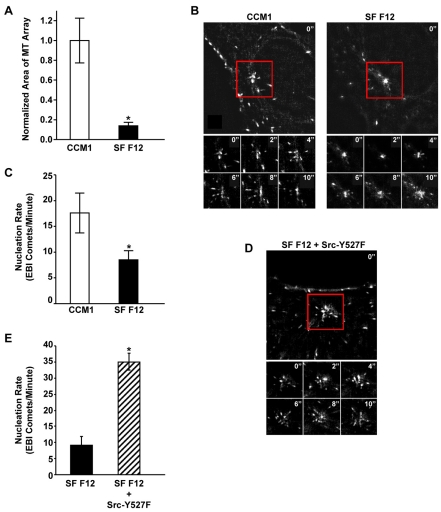
Live-cell imaging of microtubule nucleation promoted by CCM1 and Src signaling. (A) CCM1 promotes rapid microtubule regrowth in CHO-K1 cells. Mitotic cells were collected and replated onto fibronectin in either CCM1 or SF Ham's F12. Values are the normalized mean area of microtubule arrays 5 minutes post-nocodazole washout from 100 cells per condition in three independent experiments. *A statistically significant difference from the CCM1 control (P<0.05). (B-E) Time-lapse imaging was used to track newly nucleated microtubules in CHO-K1 cells stably expressing GFP-EB1. (B) Micrographs comparing EB1 comets emanating from the centrosome of representative cells replated on fibronectin in either CCM1 or SF Ham's F12. Also shown are montages of six images taken every two seconds over a 10 second period for each representative cell. (C) The average rate ± s.d. of newly nucleated microtubules calculated from ten cells per condition in three independent experiments. *A statistically significant difference from the CCM1 control (P<0.05). (D,E) Expression of the constitutively active Src-Y527F mutant promotes microtubule nucleation. (D) Micrograph of EB1 comets emanating from the centrosome of a representative Src-Y527F-transfected cell replated onto fibronectin in SF Ham's F12. Also shown is a montage of six images taken every 2 seconds over a 10 second period. (E) The average rate ± s.d. of newly nucleated microtubules from 10 cells in three independent experiments. *A statistically significant difference from the SF Ham's F12 control (P<0.05).

