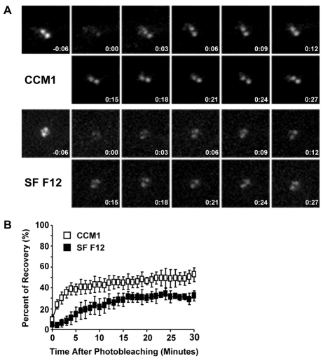
Fig. 8.
γ-Tubulin turnover at the centrosome is promoted by CCM1. (A,B) FRAP analysis using CHO-K1 cells stably expressing GFP-γ-tubulin. Mitotic cells were collected and replated onto fibronectin in either CCM1 or SF F12. (A) Representative images of pre-bleached and bleached centrosomes for each condition together with images acquired every 3 minutes post-photobleaching. (B) Quantification of the recovery of centrosomal GFP intensity. Values are the mean intensities ± s.d. from five cells per condition in three independent experiments.

