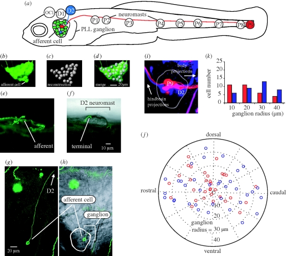
Figure 1.
(a) Afferent neurons in the posterior lateral line ganglion of 5 dpf larval zebrafish illustrating connections with D2 and P9 neuromasts (not drawn to scale). Bipolar afferents make projections that terminate on one or more neuromasts labelled OC1, D1-2 and P1-9 (Raible & Kruse 2000; Ledent 2002) as well as to the hindbrain (not shown). (b–d) Afferent cell bodies in a ganglion can be recognized and reconstructed using HUC-GFP transgenic fish. (e,g) DNA injection at the one-celled embryo stage reveals expression of a single afferent innervating the D2 neuromast. (f,h) The position of the afferent in the ganglion and its bulged termini on the neuromast is revealed by merged confocal and Nomarski microscopy images. (i,j) Backfilling dyes into D2 (blue) and P9 (red) neuromasts results in afferent cell body labelling and reveals their relative positions in the ganglion. (k) Graph showing the number of backfilled afferent neurons located in 10 µm concentric ring regions from the centre of the ganglion (n = 14 ganglia). More P9 afferent neurons reside towards the centre of the ganglion while more D2 cells reside in the outer rings of the ganglion.

