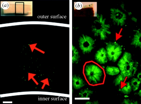
Figure 2.
Confocal images of ratite eggshells stained for DNA. (a) Confocal radial cross section of an Aepyornis eggshell, stained with SYBR Green, displaying the DNA distributed throughout the matrix: 5× objective lens, scale bar, 400 µm. Inset, orientation of confocal image. (b) Confocal inner surface of a D. robustus eggshell, stained with Hoechst dye, displaying mammillary cones (outlined) with peripherally located DNA: 40× objective lens, scale bar, 50 µm. Inset, orientation of confocal image. Red arrows, fluorescently labelled DNA.

