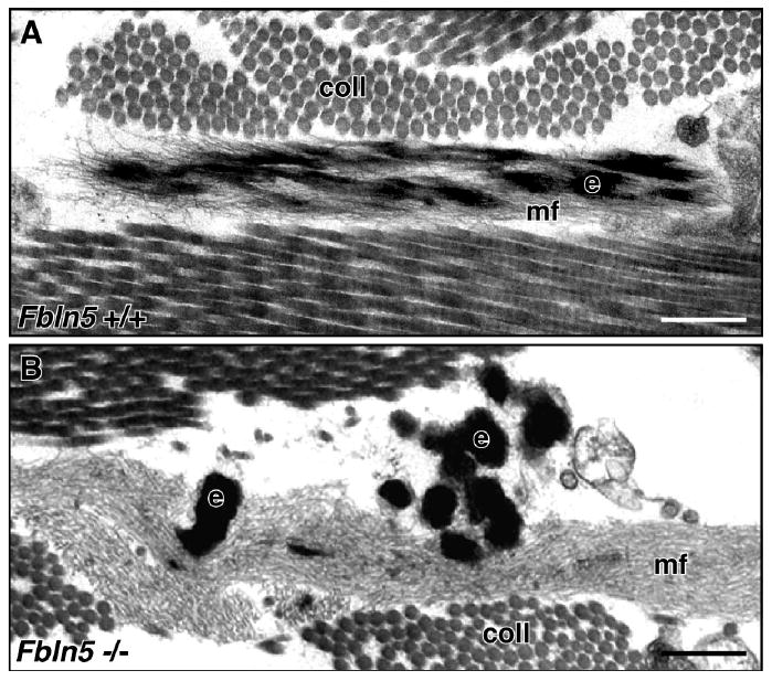
Figure 2. Electron micrographs of dermal elastic fibers in wild-type and Fbln5-null mouse skin.

(A) Elastic fibers in wild-type dermis show a core of elastin (e) integrated within a bundle of microfibrils (mf). (B) In contrast, the elastic fibers in Fbln5-null dermis are composed of large elastin aggregates (e) located outside extensive bundles of microfibrils (mf). Scale bar = 0.25 μm, coll = collagen. Adapted and used with permission from Choi et al. Matrix Biol. 28:211-220, 2009.
