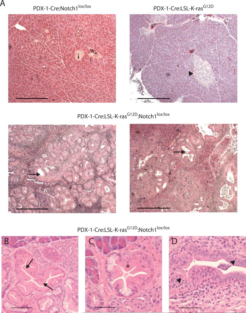
Figure 1. Histological analysis of pancreata.
(A) H&E stained sections from pancreata at 20 weeks of PDX-1-Cre:Notch1lox/lox mice (i = islet), PDX-1-Cre:LSL-K-rasG12D mice (arrowhead = tubular complexes-TC), and PDX-1-Cre:LSL-K-rasG12D:Notch1lox/lox mice (arrows = ducts exhibiting PanIN 1B-2 changes). Scale bar = 400μm. (B-D) Detailed characterization of pathology exhibited in PDX-1-Cre:LSL-K-rasG12D:Notch1lox/lox pancreata. (B) PanIN1B; papillary ductal lesions without significant loss of polarity or nuclear atypia (arrows). (C) PanIN1B transiting to PanIN2 revealing moderate nuclear atypia and a loss of polarity. (D) PanIN1B showing intraepithelial polymorphonuclear leukocytes (arrowheads). Scale bar = 200μm.

