
Figure 3. Expression of scFv EGFRvIII in stably selected hMSC cultured at various temperatures and in HEK cells.
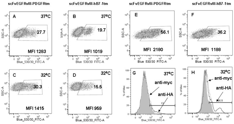
Comparison of surface expression of the scFv EGFRvIII PDGFRtm and hB7-1tm fusion in hMSC cultured at 37°C (A and B) and 32°C (C and D). Expression the scFv EGFRvIII PDGFRtm (E) and hB7-1tm fusion (F) in HEK cells cultured at 37°C 48 hours after transfection. hMSC grown at 37°C for a month show decreased scFvEGFRvIII surface expression (G), which can be “boosted” by transferring cells to 32°C (H) as detected by anti-HA antibodies by flow cytometry.