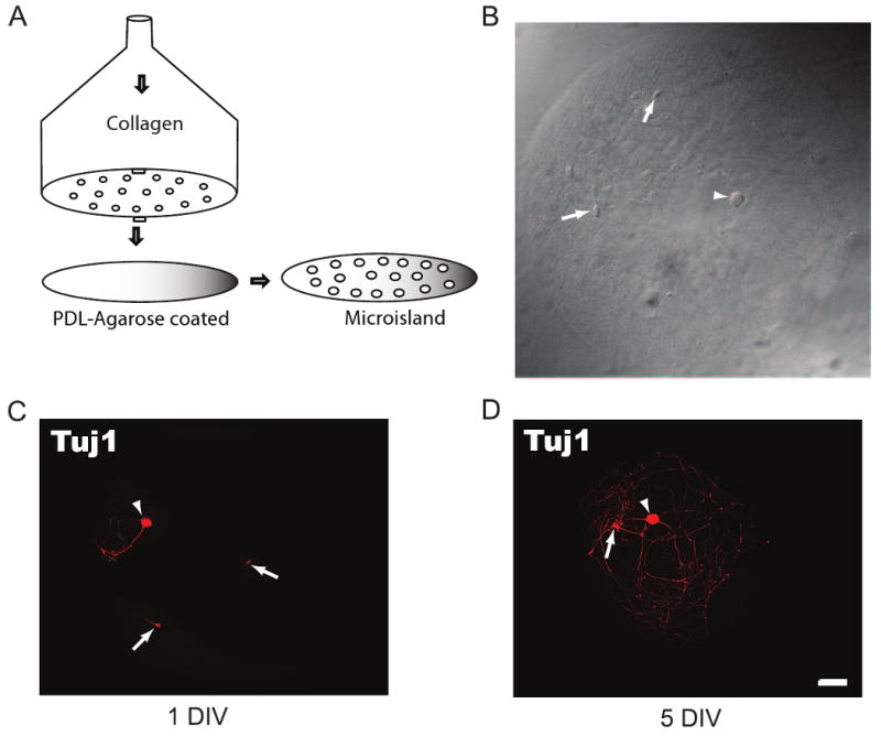
Fig. 1.

DRG/dorsal horn neuron microisland assay system. (A) Schematic of the homemade device for making microisland coverslips showing the perforations through which collagen is pushed out by air to generate a matrix of dots on a PDL-agarose pre-coated coverslip. (B) Example of a microisland with a DRG (Arrowhead) and two dorsal horn neurons (Arrows). (C, D) DRG (Arrowheads) and dorsal horn neurons (Arrows) at day 1 (C) and 5 (D) in vitro are positive for the neuronal marker Tuj1. Neurons avoid the nonpermissive agarose and are confined to the permissive substrate (collagen/cortical astrocytes). Note that a DRG neuron is easily identified by its size difference from the dorsal horn neuron. Scale bar is 50μm.
