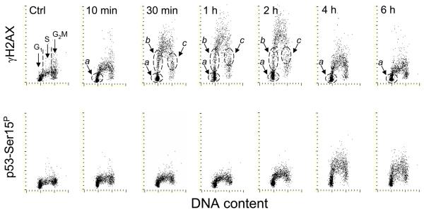
Fig. 2. Phosphorylation of H2AX on Ser139 and p53 on Ser15 after exposure of A549 cells to 50 J/m2 of UV-B light.
The cells growing on slides were irradiated with UV light, as described in the legend to Fig 1, and then were cultured for different time intervals as marked. As in Fig. 1, subpopulations a, b, c and d were identified.

