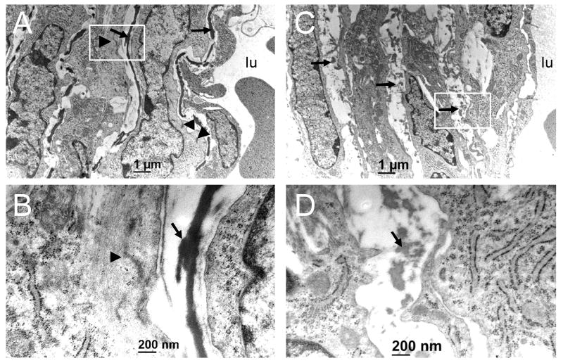
Figure 2. The loss of Dicer in VSM causes structural abnormalities.
Cross-sections of Dfl/wt (A,B) and SMC-Dicer KO (C,D) dorsal aorta from E15.5 embryos. The white boxed region in panels A and C (12,000x) are depicted in panels B and D under higher magnification (60,000x). Arrows denote elastic lamellae and arrowheads point to dense plaques and myofilament arrays seen most prevalently in SMC of wildtype aorta. lu, lumen.

