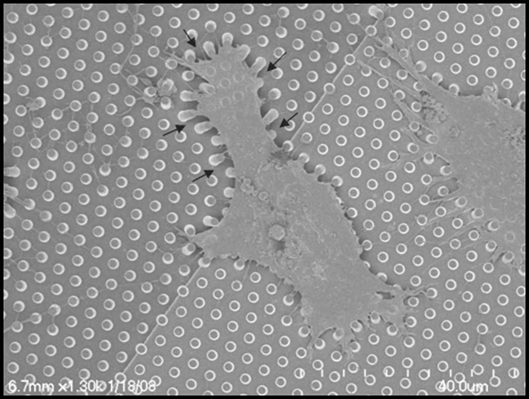
Figure 6.
SEM image of an immortalized mesenchymal stem cell attached to a hexagonal array of posts with a constant diameter of 1 mm, pitch of 2 mm and two different heights of 6.6 and 3.9 mm, which causes a change in rigidity of the substrate. The cell is more spread on the rigid part (right part) than the softer part (left part). Also, on the softer area the posts are bent to a greater extent compared to the rigid posts. The arrows indicate the direction of pillar deflection.

