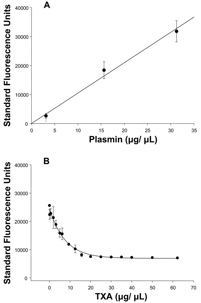
Figure 1.
(A) Fluorescence emission of the plasmin specific substrate (6.25µg/mL), reflective of PLact, increased with increasing concentrations of plasmin (0–31.25 µg/mL) in a linear concentration dependent manner (n=3, plotted values are mean±SEM; linear regression, y(x) = 1048.8*x, r2 = 0.996, p = 0.002).
(B) Fluorescence emission of the plasmin specific substrate (6.25µg/mL), reflective of PLact, in the presence of plasmin (31.25µg/mL) and control porcine plasma (1:32) decreased in response to increasing concentrations of TXA (0–62.2mg/mL)in a classic logarithmic concentration dependent manner 13 (n=3, plotted values are mean±SEM, regression, y(x) = 23280*e−0.063*x, r2=0.964, p<0.001).

